
What is 3D Echocardiography?
3D Echo uses advanced technology to create three-dimensional real-time images of the heart. With GE Vivid E95, excellent spatial resolution is achieved allowing accurate valve, cavity and structure assessment.
Three-dimensional visualization of cardiac structures
More accurate valve assessment
Improved preoperative planning
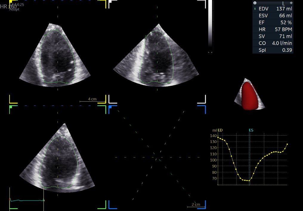
GE Vivid E95: Leading 3D Imaging Technology
The GE Vivid E95 uses a matrix array probe that simultaneously records thousands of data points, creating live 3D/4D images of the heart in real time. The anatomical detail achieved far surpasses conventional 2D imaging, revealing pathologies that would otherwise go undetected, even in difficult imaging cases.
The clinical value of 3D Echo is particularly significant in valve disease. For example, in mitral valve regurgitation, 3D Echo precisely reveals the mechanism (prolapse, chordal rupture, annular dilatation) and enables the mapping that the surgeon needs for repair. Automated algorithms (Auto EF 3D) calculate cardiac chamber volumes with high reproducibility, minimising measurement error.
Combined with Strain Analysis (GLS), 3D Echo detects early myocardial dysfunction before ejection fraction falls — a critical finding for patients receiving cardiotoxic chemotherapy. The examination requires no special preparation and is performed as an extension of the standard heart triplex.
When is 3D Echo needed?
Valve Diseases
Accurate valve assessment
Preoperative Planning
Before valve surgery
Congenital Heart Disease
Anomaly investigation
Cavity Masses
Ejection fraction calculation
Cardiomyopathies
Myocardial structure assessment
Accurate Diagnosis
When 3D analysis is required
How is 3D Echo performed?
3D Echo is performed like classic heart triplex, using a special 3D probe that collects three-dimensional heart images in real-time through GE Vivid E95.
- Patient lies on examination bed
- Gel and 3D probe are placed on chest
- Three-dimensional images are acquired in real-time
Duration
25-35 minutes
3D Echo Advantages
Superior Analysis
Three-dimensional structure visualization
Accurate Assessment
Improved valve estimation
Preoperative Planning
Surgical guidance
GE Vivid E95
Top technology worldwide